NCERT Solutions for Class 11th Biology Chapter 20: Locomotion and Movement
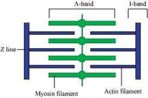
Question 1: Draw the diagram of a sarcomere of skeletal muscle showing different regions.
Answer The diagrammatic representation of a sarcomere is as follows:
Question 2:Define sliding filament theory of muscle contraction.
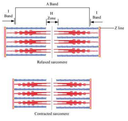
Answer The sliding filament theory explains the process of muscle contraction during which the thin filaments slide over the thick filaments, which shortens the myofibril. Each muscle fibre has an alternate light and dark band, which contains a special contractile protein, called actin and myosin respectively. Actin is a thin contractile protein present in the light band and is known as the I-band, whereas myosin is a thick contractile protein present in the dark band and is known as the A-band. There is an elastic fibre called z line that bisects each I-band. The thin filament is firmly anchored to the z line. The central part of the thick filament that is not overlapped by the thin filament is known as the H-zone. During muscle contraction, the myosin heads or cross bridges come in close contact with the thin filaments. As a result, the thin filaments are pulled towards the middle of the sarcomere. The Z line attached to the actin filaments is also pulled leading to the shortening of the sarcomere. Hence, the length of the band remains constant as its original length and the I-band shortens and the H-zone disappears.
Question 3: Describe the important steps in muscle contraction.
Answer During skeletal muscle contraction, the thick filament slides over the thin filament by a repeated binding and releases myosin along the filament. This whole process occurs in a sequential manner.
Step 1: Muscle contraction is initiated by signals that travel along the axon and reach the neuromuscular junction or motor end plate. Neuromuscular junction is a junction between a neuron and the sarcolemma of the muscle fibre. As a result, Acetylcholine (a neurotransmitter) is released into the synaptic cleft by generating an action potential in sarcolemma.
Step 2: The generation of this action potential releases calcium ions from the sarcoplasmic reticulum in the sarcoplasm
Step 3: The increased calcium ions in the sarcoplasm leads to the activation of actin sites. Calcium ions bind to the troponin on actin filaments and remove the tropomyosin, wrapped around actin filaments. Hence, active actin sites are exposed and this allows myosin heads to attach to this site.
Step 4: In this stage, the myosin head attaches to the exposed site of actin and forms cross bridges by utilizing energy from ATP hydrolysis. The actin filaments are pulled. As a result, the H-zone reduces. It is at this stage that the contraction of the muscle occurs.
Step 5: After muscle contraction, the myosin head pulls the actin filament and releases ADP along with inorganic phosphate. ATP molecules bind and detach myosin and the cross bridges are broken.
Stage 6: This process of formation and breaking down of cross bridges continues until there is a drop in the stimulus, which causes an increase in calcium. As a result, the concentration of calcium ions decreases, thereby masking the actin filaments and leading to muscle relaxation.
Question 4: Write true or false. If false change the statement so that it is true.
(a) Actin is present in thin filament
(b) H-zone of striated muscle fibre represents both thick and thin filaments.
(c) Human skeleton has 206 bones.
(d) There are 11 pairs of ribs in man.
(e) Sternum is present on the ventral side of the body.
Answer (a) Answer: True
(b) Answer: False
H -zone of striated muscle fibre is the central part of the thick filament that is not overlapped by the thin filament.
(c) Answer: True
(d) Answer: False
There are 12 pairs of ribs in a man.
(e) Answer: True
Question 5: Write the difference between:
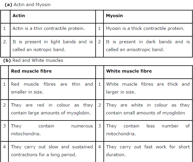
(a) Actin and Myosin
(b) Red and White muscles
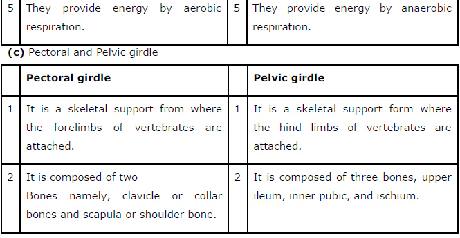
(c) Pectoral and Pelvic girdle
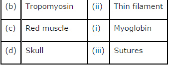
Answer
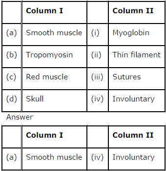
Question 6:
Match Column I with Column II :
Question 7: What are the different types of movements exhibited by the cells of human body?
Answer Movement is a characteristic feature of living organisms. The different types of movement exhibited by cells of the human body are:
• Amoeboid movement: Leucocytes present in the blood show amoeboid movement. During tissue damage, these blood cells move from the circulatory system towards the injury site to initiate an immune response.
• Ciliary movement: Reproductive cells such as sperms and ova show ciliary movement. The passage of ova through the fallopian tube towards the uterus is facilitated by this movement.
• Muscular movement: Muscle cells show muscular movement.
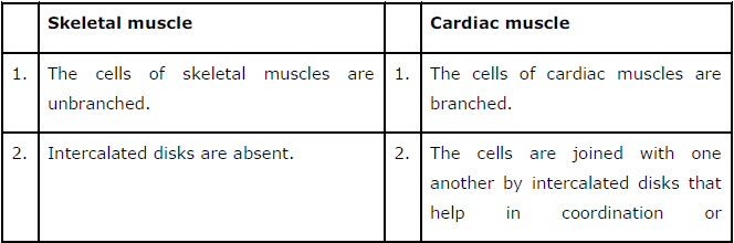
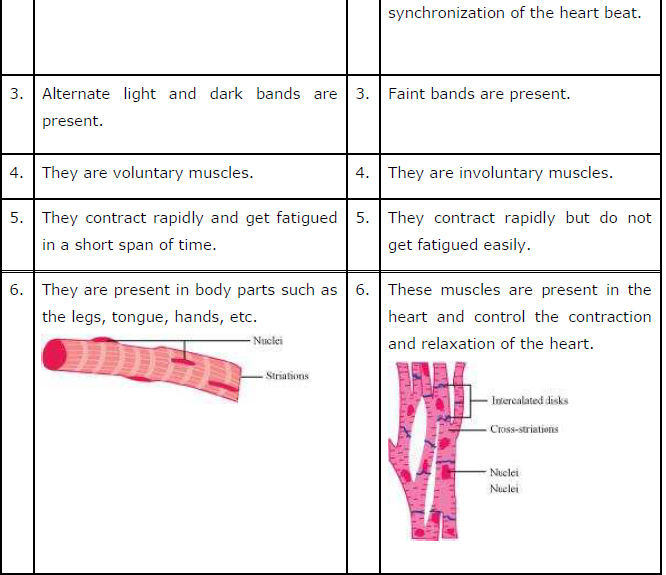
Question 8: How do you distinguish between a skeletal muscle and a cardiac muscle?
Answer
Question 9: Name the type of joint between the following:-
(a) atlas/axis
(b) carpal/metacarpal of thumb
(c) between phalanges
(d) femur/acetabulum
(e) between cranial bones
(f) between pubic bones in the pelvic girdle
Answer(a) atlas/axis: Pivotal joint
(b) carpal/metacarpal of thumb: Saddle joint
(c) between phalanges: Hinge joint
(d) femur/acetabulum: Ball and socket joint
(e) between cranial bones: Fibrous joint
(f) between pubic bones in the pelvic girdle: Ball and socket joint
Question 10: Fill in the blank spaces:
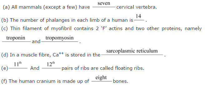
(a) All mammals (except a few) have __________ cervical vertebra.
(b) The number of phalanges in each limb of human is __________
(c) Thin filament of myofibril contains 2 ‘F’ actins and two other proteins namely _________ and __________.
(d) In a muscle fibre Ca++ is stored in __________
(e) __________ and __________ pairs of ribs are called floating ribs.
(f) The human cranium is made of __________ bones.
Answer
Go Back to NCERT Solutions Biology Page Physics Chemistry Maths
To start your test
register with us:
To start
your test, registered user must login with their User Name & Password: